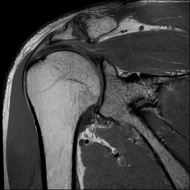
photo credit: radiopaedia.org
Magnetic resonance imaging of musculoskeletal pain has long been known to find various pathologies, including herniated disks, tendon tears, and spinal stenosis, which may or may not contribute to an individual’s symptoms. These spinal and extremity findings are common in pain free individuals especially over the age of 30 years old. As MRI machine technology continues to improve its’ ability to detect musculoskeletal changes clinicians must ensure a patient’s history and clinical examination matches with the images’ findings. An interesting study was recently conducted examining not only the involved but also the uninvolved joint of patient’s with shoulder pain to determine which MRI findings contributed to a patient’s symptoms.
A recent research article in the Journal of Shoulder and Elbow Surgery reported on the frequency and severity of musculoskeletal structural pathologies among patients with shoulder pain (Goncalves Carreto et al. 2019). Authors recruited 123 patients with unilateral shoulder pain. Participants were excluded if they had a history of upper limb fracture, repeated dislocations, or signs of adhesive capsulitis. Each individual underwent a MRI of his or her involved and uninvolved shoulder with images being read by both an orthopedic surgeon and musculoskeletal radiologist. As expected abnormal MRI findings were frequent, but surprisingly were found in both the symptomatic and pain free shoulder. The majority of pathologies were found in equal frequency between shoulders but some findings including full thickness rotator cuff tears and osteoarthritis were higher (10% more frequent) in the involved shoulder. The researchers reiterated prior researchers advice to correlate these images’ findings with a patient’s age, history, and clinical examination.

