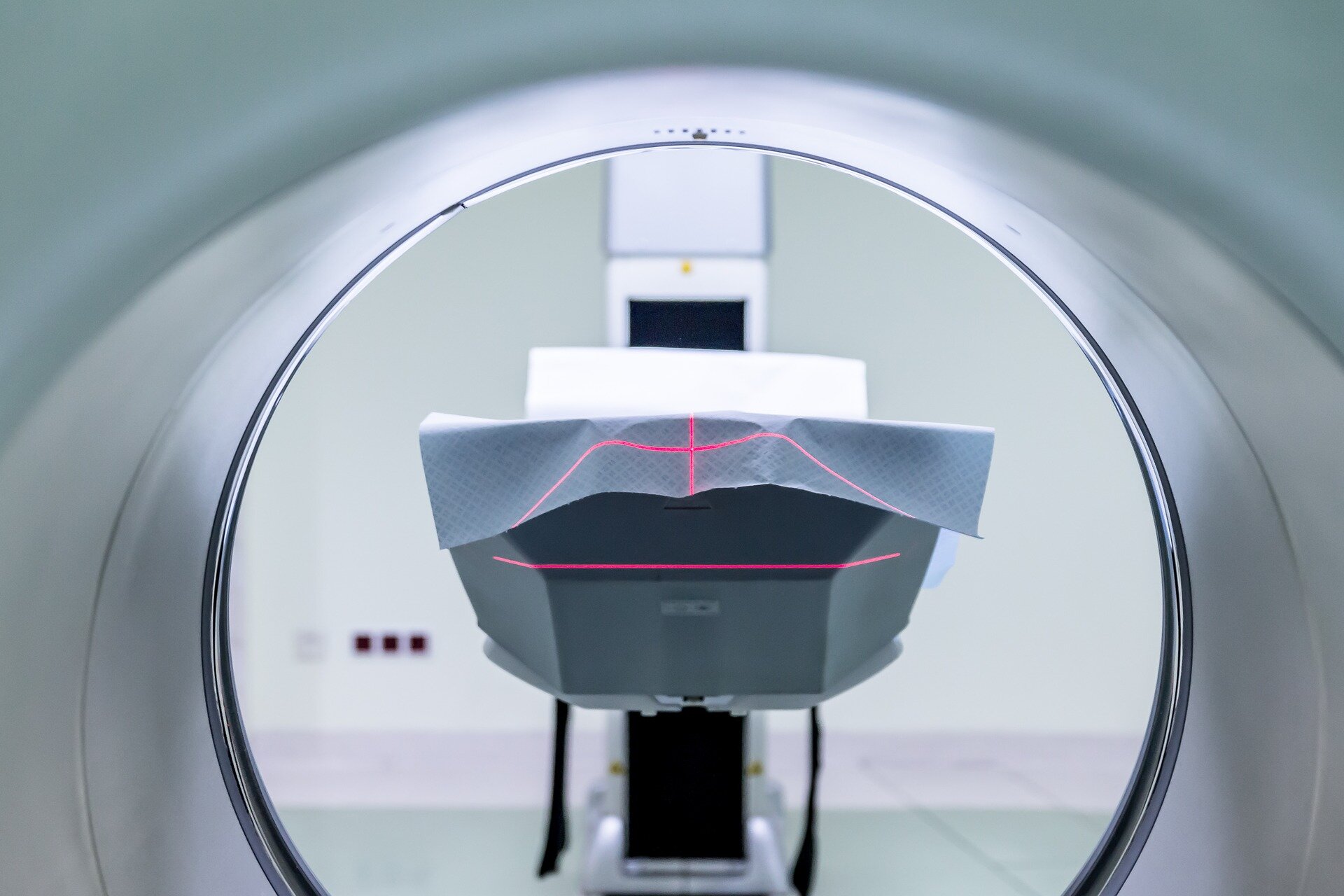
The old saying that you can’t unring a bell is true in imaging for musculoskeletal conditions. Most patients cannot unsee their MRI results despite improvements in their symptoms and activity level with Physical Therapy treatments. Once terms such as bulging or degeneration have been utilized to describe their spines they never regain full confidence in this region of the body. Previous studies in patients with low back pain have found those who were given the results of their baseline images had less recovery than their peers who were blinded to the results of their imaging. Pooled results of the academic research consistently do not find any added benefit of early MRI imaging in patients with low back pain.
Many of the structural changes found on MRI of patients with low back pain were likely present when they were pain free and will continue to be present or improve when patients become pain free again after Physical Therapy treatments. In a classic study, Carragee and colleagues recruited 200 participants without low back pain and gave them a baseline lumbar spine MRI (The Spine Journal. 2006). Then these participants were followed for up to 5 years and asked to return to the authors for a 2nd lumbar MRI if they were having significant low back pain or leg pain symptoms (>6/10 NPRS). Close to 25% patients returned for the 2nd MRI and of these 84% showed no change or an improvement in their MRI scans. Changes in the other 16% of these patients were attributed to age related changes and time, but were not consistent with their new low back pain symptoms. Authors concluded structural changes on MRI did not predict onset of low back pain symptoms.
A recent study in the journal Spine supports these earlier findings by Carragee et al. Simo and colleagues were interested in determining if early degenerative disc disease changes were associated with future pain, disability, or MRI findings (Spine. 2020). Authors initially examined 75 patients with low back pain with a clinical exam and MRI. These individuals were then contacted at a 30 year follow up to determine how their low back pain symptoms changed since their baseline examination. They reported the severity of disc degeneration at baseline did not predict who would have the most back pain or disability at the 30 year follow up. Conversely, patients with early degeneration at baseline did have greater amounts of degeneration at long term follow up showing the continued lack of correlation between imaging findings and a patient’s clinical presentation or activity level.
Patients with low back pain are encouraged to view their imaging findings as an opportunity to rule out serious conditions not as an accurate predictor of their future back pain or ability levels.
Click Here to learn which low back pain exercises are best for your symptoms

